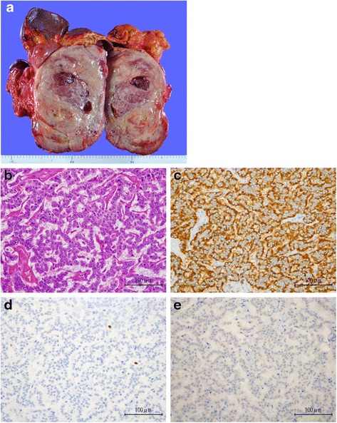
Fig. 3.

a Excised insulinoma, measuring 18.0 × 13.0 × 12.0 cm. b Hematoxylin-eosin staining of the insulinoma (×400). c Tumor cells are positive in the immunohistochemical staining for chromograin A (×400). d Ki67 is 4.3 % (×400). e Tumor cells are focally positive for insulin (×400)
