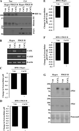
Fig. 9.

HIF-1α activation supports extracellular acidosis. A: immunoblot analysis of HIF-1α (arrow) in the nuclear and cytoplasmic fractions from vector control (Hygro) or PDGF-B BPH-1 cells with vehicle only or 100 μM CoCl2 treatment. Histone H1 and PTEN were used as loading controls for the nuclear and cytoplasmic fractions, respectively. Upon treatment of vector (Hygro) or PDGF-B BPH-1 cells with CoCl2, CA mRNA expression (RT-PCR) (B), changes in extracellular pH (C–F), and matriptase activation (G) were monitored. Values represent the mean ± SD. *P < 0.05. Ponceau S was used to evaluate equal gel loading.
