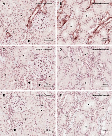
Fig. 10.

CaSR detection by proximity ligation assay in rat kidney. Photomicrographs of proximity ligation assay using the CaSR antibody pair N-term1/N-term6 in outer (A) and inner (B) cortical kidney sections. Photomicrographs of proximity ligation assay using the CaSR antibody pair N-term1/N-term2 in outer (C) and inner (D) cortical kidney sections are shown. Photomicrographs are also shown of proximity ligation assay using the CaSR antibody pair N-term2/C-term1 in outer (E) and inner (F) cortical kidney sections. Positive staining corresponds to immunoperoxidase staining (brown dots). Asterisks indicate PT. Scale bar = 50 μm.
