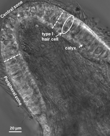
Fig. 1.

Differential interference contrast (DIC) image of a transverse slice cut from the midregion of a postnatal day (P)22 gerbil crista. White dashed lines show the boundary between the central zone (CZ) and peripheral zone (PZ). Long arrow indicates a typical type I hair cell and short arrow a cup-shaped calyx terminal. Patch electrodes targeted the unmyelinated basal surface of the outer face of calyx terminals. Hair cells are shorter and less densely packed in the CZ. Stereocilia extend from the cuticular plates of many hair cells, and myelinated axons can be seen coursing through the stroma.
