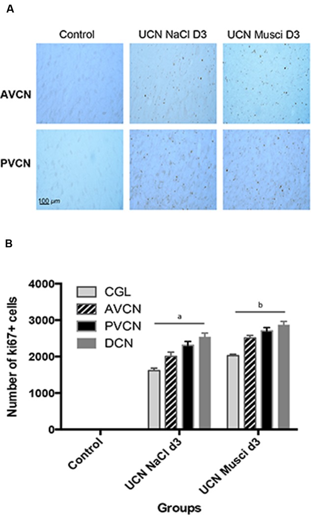
FIGURE 3.
Ki67 immunostaining was used to confirm BrdU cell proliferation. (A) Photomicrographs showing Ki67 immunostainings in the deafferented AVCN and PVCN of control cats and cats infused with NaCl or Muscimol immediately after UCN and at sacrifice (D3). Scale bar: 100 μm. (B) Quantitative evaluation. Histograms comparing the mean values (±SEM) of Ki67-immunopositive cells in the deafferented CN 3 days after UCN in control cats or cats submitted to NaCl or Muscimol infusion using ANOVA followed by the Scheffé test (p < 0.0001, see Table 2). Data from both sides of control animals were pooled for direct comparison with the subgroups of lesioned cats. Different letters indicate significant differences between groups of animals in each CN: a: significantly different from control and UCN Muscimol groups; b: significantly different from control and UCN NaCl groups. Only values recorded on the lesioned side are illustrated. n = 4 animals per group.

