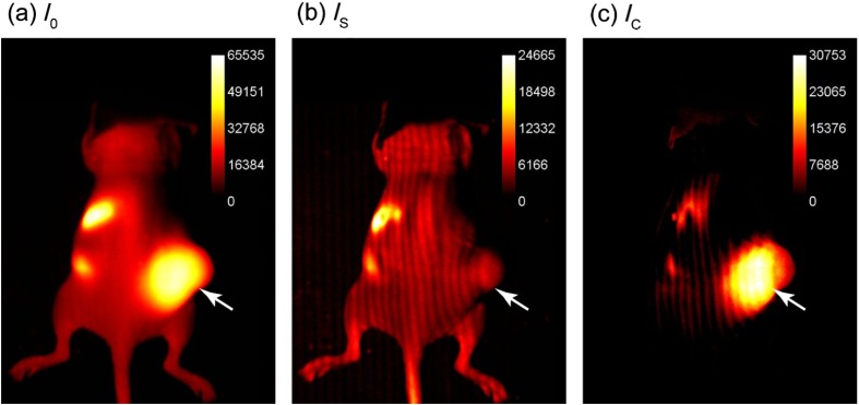
Fig. 2.
Demonstration of SIFMI process with subcutaneous tumor xenograft model and NIR fluorescent molecular probe with high affinity for multiple myeloma cancer cells in solid tumor (arrow). (a) Planar fluorescence uniform illumination equivalent image () reconstructed using the sum of the projected light patterns [Eq. (2)]. (b) Surface signal image () from the modulated signals [Eq. (3)]. (c) Subsurface, diffuse signal () according to a modified Eq. (1).

