
Figure 5.
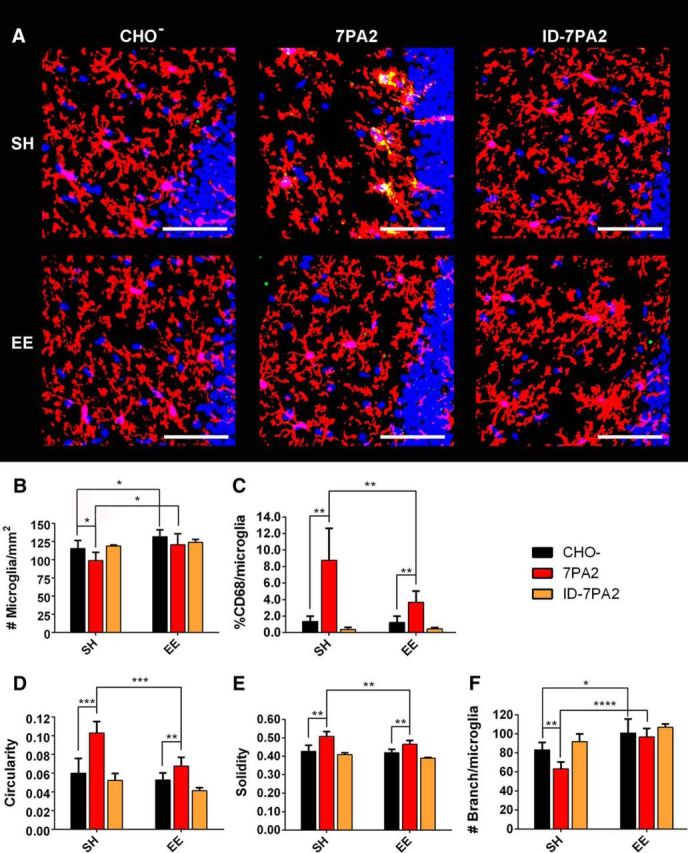
Oligomeric Aβ induces changes in microglial density and morphology in SH mice, and EE prevents such changes. A, Representative images from SH and EE mice receiving 7PA2 CM, CHO− CM, or immunodepleted 7PA2 CM (ID-7PA2) show strong microglial alteration in SH from 7PA2 CM, as reflected by microglial morphology (microglia labeled with P2ry12 antibody, red) and high level of CD68 (green). DAPI (blue) demonstrates that all images are from consistent DG area. EE potently neutralizes microglial alteration. Removing oAβ from 7PA2 CM also successfully abolishes microglial alteration. Scale bar, 50 μm. B–F, Quantification: 7PA2 CM significantly decreases DG microglial density, significantly increases % CD68/microglia, circularity, and solidity, and decreases #branch/microglia in SH mice. EE mice upon 7PA2 CM exposure have significantly higher microglial density than that of SH mice, and show significantly different morphological features: smaller value of % CD68/microglia, circularity, and solidity, as well as higher #branch/microglia. EE mice receiving CHO− CM show significantly higher DG microglial density and higher #branch/microglia than CHO− CM-receiving SH mice, just as in naive mice. ID-7PA2 CM shows no difference from CHO− CM in both SH and EE mice. *p < 0.05. **p < 0.01. ***p < 0.001. ****p < 0.0001. N = 6. Quantitative data are mean ± SEM.