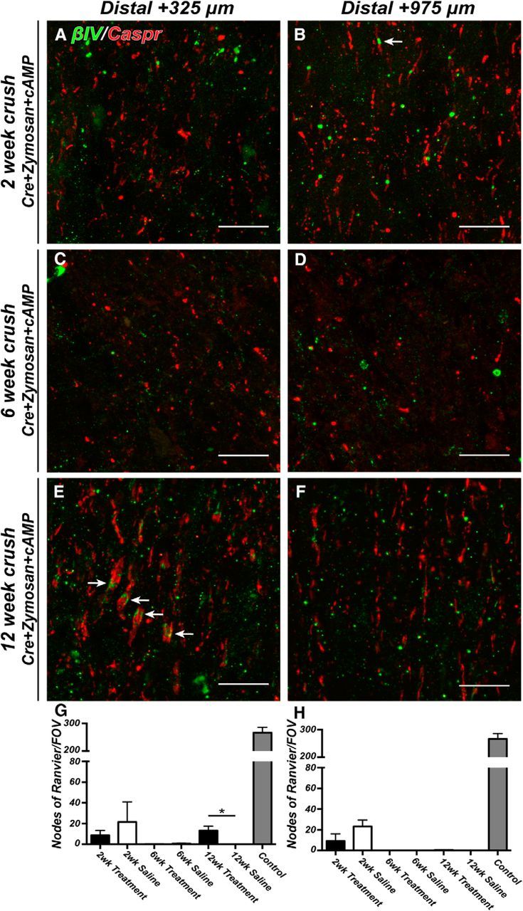
Figure 6.

Nodes of Ranvier are reassembled in regenerating CNS axons distal to the crush site. A–F, Representative images of optic nerves immunostained for βIV spectrin (green, arrows) and Caspr (red) distal to the crush (A, C, E: 325 μm; B, D, F: 925 μm). Optic nerves were examined at 2, 6, and 12 weeks after crush. G, H, The ratio of nodes of Ranvier per FOV in crushed optic nerve to uninjured control optic nerve at 2, 6, and 12 weeks after crush. Measurements were made distal to the crush site (+325 and +975 μm). Treatment = Cre+zymosan+cAMP. Scale bars: A–F, 10 μm. *p < 0.05 (unpaired t test with Mann–Whitney post test).
