Figure 9.
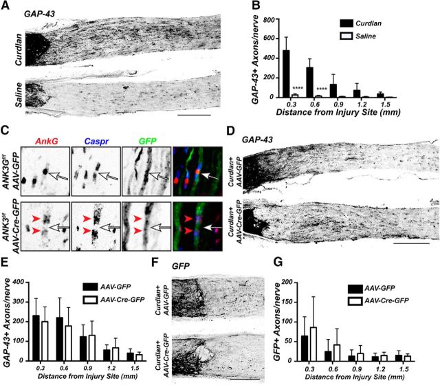
AnkG is not required for axon regeneration. A, GAP-43 immunostatining of optic nerve after intravitreal injection of β-1,3-glucan (Curdlan) or saline, 2 weeks after optic nerve crush. B, Quantification of GAP-43-labeled axons in mice administered either Curdlan or saline 2 weeks after optic nerve crush. C, Immunostaining of Ank3f/f mouse optic nerves after intravitreal injection of either AAV-GFP or AAV-Cre-GFP using antibodies against ankG (red), Caspr (blue), and GFP (green). Arrows indicate nodes of Ranvier. Red arrowheads indicate paranodal ankG in the AAV-Cre-GFP injected mice. D, F, Optic nerves from Ank3f/f mice treated with Curdlan and either AAV-GFP or AAV-Cre-GFP and immunostained with GAP-43 (D) or GFP (F) 2 weeks after optic nerve crush. E, G, Quantification of GAP-43 (E) or GFP (G) positive axons in regenerating axons in Ank3f/f mice administered curdlan and either AAV-GFP or AAV-Cre-GFP 2 weeks after optic nerve crush. Scale bars: A, D, 300 μm; F, 200 μm. ****p < 0.0001 (unpaired t test with Mann–Whitney post test).

