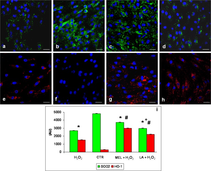
Fig. 3.
Immunofluorescence analyses for superoxide dismutase 2—SOD2 (a–d) and heme oxygenase-1—HO-1 (e–h) in L6 myotubes incubated with hydrogen peroxide—H2O2 (a, e), control—CTR (b, f), pre-treated with melatonin and then incubated in hydrogen peroxide—MEL + H2O2 (c, g) and pre-treated with LA and then incubated in hydrogen peroxide—LA+ H2O2 (d, h). Nuclei were stained with DAPI. Scale bar = 20 μm. The graph (i) summarizes the quantitative analysis of immunopositivities. * p < 0.05 vs control cells, #p < 0.05 vs H2O2-treated cells and +p < 0.05 vs pre-treated with melatonin and then incubated in hydrogen peroxide

