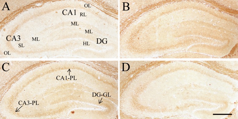
Fig. 3.
Effect of age or DNJ on insulin receptor (InsR) immunohistochemical staining in the dorsal hippocampus of the SAMP8 mice. Representative low-magnification photos in the O-con (a), Y-con (b), HD-DNJ (c), and LD-DNJ (d) groups are shown. The InsRs were densely expressed in the layers containing cell bodies, i.e., DG-GL, CA1-PL, and CA3-PL. The old controls had lower InsR immunoreactivity than the young controls, which was alleviated by the HD-DNJ treatment. DG dentate gyrus, GL granule cell layer, PL pyramidal cell layer. Scale bar = 400 μm

