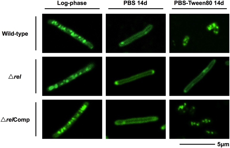
FIGURE 3.

Lipid body staining images of M. smegmatis Δrel in comparison with the wild-type and complemented strain. Fluorescent probe stained culture samples of M. smegmatis Δrel grown in rich broth (log-phase) and starved in PBS or PBS-Tween80 for 14 days in comparison with wild-type M. smegmatis and M. smegmatis Δrel complemented strain (ΔrelComp) are shown. Staining was done with Nile red (green) to visualize intracellular lipid bodies. Nile red stains predominantly intracellular lipid inclusions. Representative fields were shown.
