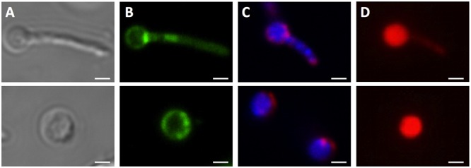
FIGURE 4.

Unusual cell shapes in PBS-starved cultures of M. smegmatis Δrel. Typical examples of unusual ‘ball-like’ cell shapes (∼60%) observed in 14-day PBS-starved M. smegmatis Δrel cultures are shown. Cells carried ball-like appendices are shown in the upper panels, and ball-shaped cells are shown in the lower panels. (A) Differential interference contrast images to visualize cell shapes. (B) Nile red (green) stained samples to visualize lipid bodies. (C) DAPI (blue) and FM4-64 (red) double stained samples to visualize DNA and membrane, respectively. (D) Propidium iodide (red) staining was done to show breakdown of the permeability barrier of the ball-like structures. These structures were observed under phase contrast but not in acid-fast stained samples, suggesting that these structures may be cell wall defective. Representative fields were shown. White bar = 1 μm.
