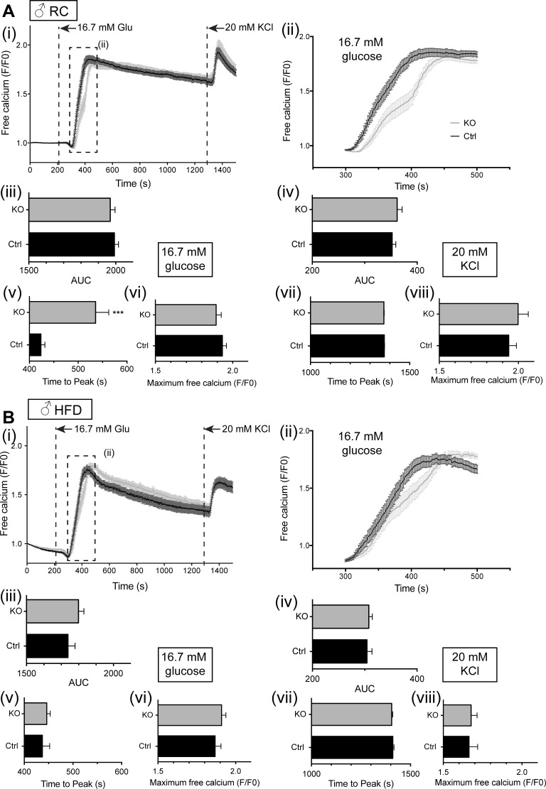
Fig. 10.
Effect of Vps13c deletion on calcium signaling in vitro in male mouse islets. Isolated islets from male mice (20–23 wk of age), maintained on RC (A) or HFD (B) were loaded with Fluo 2 and incubated in Krebs-Ringer solution containing 3 mM glucose (3 mM Glu) for 45 min. Dye-loaded islets (3–7 per field of view) were imaged on a spinning disk confocal microscope for 2 min in 3 mM glucose, as described in materials and methods. A perifusion system was used to allow subsequent imaging of the islets in 16.7 mM Glu for 18 min, followed by 20 mM KCl for 5 min. Individual traces from each islet were then averaged to give one trace per islet, which was then pooled with the other islets. (i), mean free Ca2+ (normalized to initial fluorescence; F/F0); (ii), inset from (i), mean free Ca2+ measured between 300 and 500 s, showing the effect of stimulation with 16.7 mM glucose; (iii), AUC analysis for high glucose stimulation; (iv), AUC analysis for KCl stimulation; (v), time to maximum peak value from stimulation with glucose; (vi), maximum peak value (F/F0) from stimulation with glucose; (vii), time to maximum peak value from stimulation with KCl; (viii), maximum peak value (F/F0) from stimulation with KCl; n = 3–5 mice per genotype. Number of islets used: n = male RC 31–38 islets from 3 mice; male HFD mice n = 41–46 islets from 4 mice. *P < 0.5, **P < 0.01, ***P < 0.001, unpaired Student's t-test.

