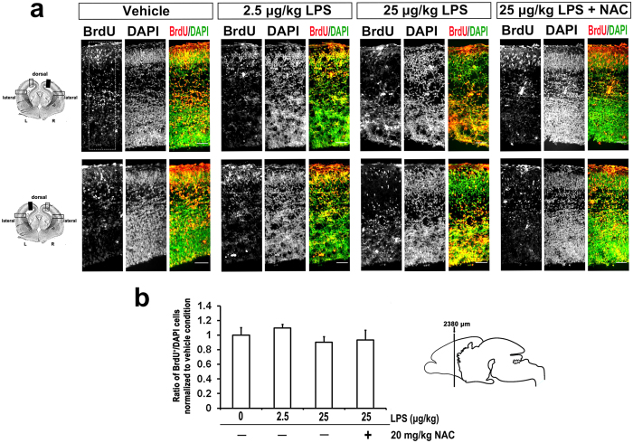
Figure 4. Maternal LPS and NAC exposure had no effects on embryonic neurogenesis.
BrdU (50 mg/kg/day) were injected intradermally from GD 15 to GD 18 after exposure to LPS. Animals were sacrificed at GD 18 and immunohistochemistry was followed. Coronal sections of indicated GD 18 rat embryonic brains were immunostained for BrdU and DAPI. Merged image is composed of BrdU (red) and DAPI (green). (a) Representative images of the sections at cortical dorsal area from three independent experiments are shown. BrdU+ cells were displayed closely to ventricles in high doses of LPS treated group compared to vehicle exposed group, which BrdU+ neurons were localized mostly in cortical plate. The effects of NAC returned the abnormal laminar characteristic to control levels. Scale bar, 50 μm. (b) displays the quantitative results of BrdU+ and DAPI+ cells in the indicated dorsal area of cortex (dotted square in (a)). Four embryo brains were randomly selected from at least three different pregnant rats. In each brain, two coronal sections near the site around 2380 μm from the front of olfactory bulb were analyzed. Ratio of BrdU+/DAPI in each condition was normalized to the PBS-exposed group. LPS treatment had no effects on the neurogenesis, determined by ANOVA followed by Tukey-Kramer Multiple Comparisons Test.

