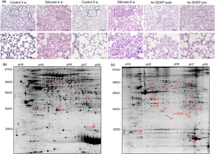
Figure 1. The pathological observation and 2-DE gel electrophoresis in rats.
(a) Representative micrographs for H.E. staining in the lung from different groups as indicated. Scale bar = 200 μm and 50 μm. (b) Soluble protein extracts were analyzed in first dimension (pH 3–10 NL IPG, 24 cm); second dimension was performed on a vertical slab (13%T) gel. Protein detection was achieved by using colloidal Coomassie staining. (c) The 2-DE partern of insoluble protein extracts. Numbering refers to differentially-represented protein spots in the silicosis model, which were then excised, digested and identified by MS procedures as reported in Table S1–4.

