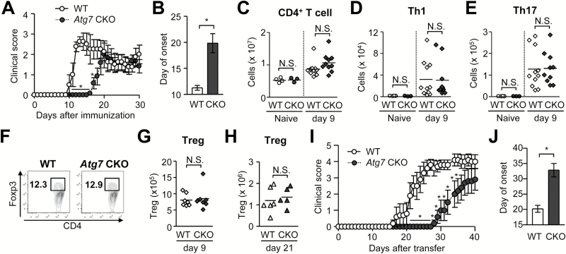
Fig. 1.
Delayed onset and mild EAE in Atg7 CKO mice. (A, B) Time-course of EAE score (A) and average day of EAE onset (B). WT (n = 6). Atg7 CKO (n = 4). Error bars denote mean ± SD. *p < 0.05. (C–E) Numbers of CD4+ T cell (C), Th1 (D), Th17 (E) cells in DLNs on days 0 and 9 after EAE induction. (F) Representative flow cytometry charts of CD3-positive pre-gated cells in DLNs on day 9. (G and H) Numbers of Tregs (Foxp3+CD4+) in DLNs on day 9 (G) or day 21 (H). (I and J) Clinical score (I) and onset (J) of passive EAE. CD4+ T cells obtained from 2D2 mice were cultured with plate-coated anti-CD3 and anti-CD28 antibodies (5 μg ml−1 each) in Th17-polarizing condition for 5 days and adoptively transferred to sub-lethally irradiated WT and Atg7 CKO recipients. n = 5 per group. One data point reflects a result from one mouse. Horizontal lines denote average values. Data are representative of two independent experiments. N.S.: not significant.

