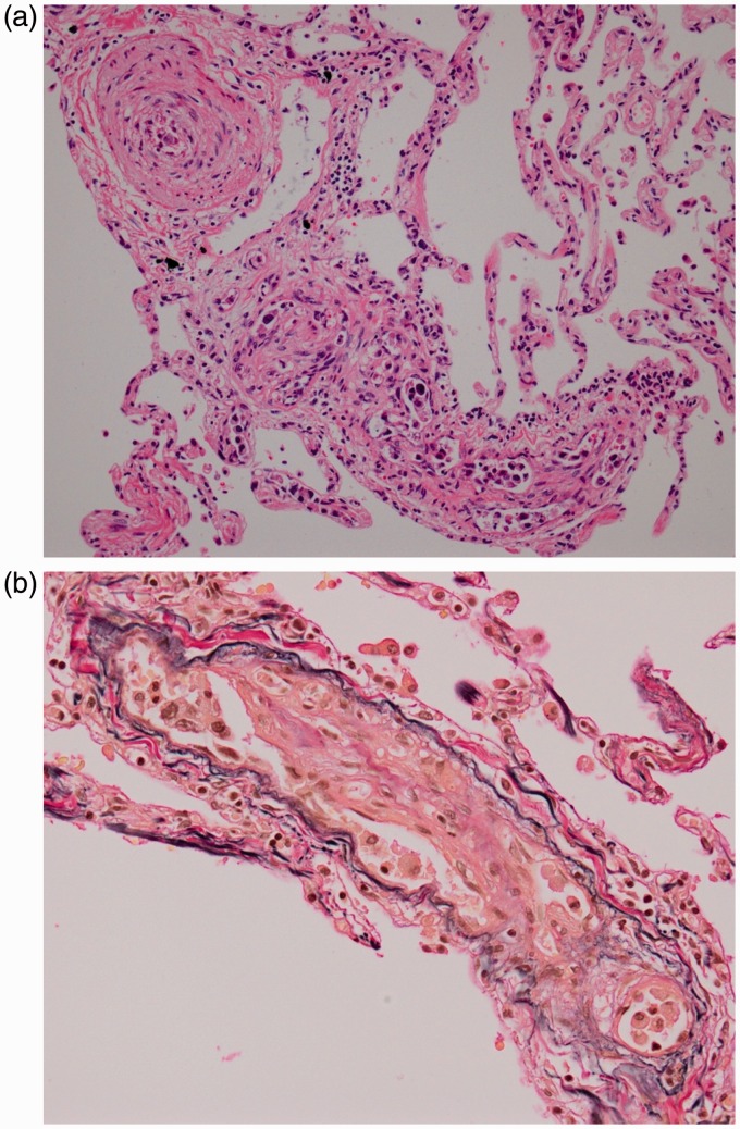
Fig. 3.
(a) H&E staining of the lung showing adenocarcinoma cells embolizing small pulmonary arteries with fibrocellular intimal proliferation. No hemorrhage or infiltration of inflammatory cells can be seen. (b) Elastica van Gieson staining showing fibrous thickening and fibrocellular intimal proliferation of endothelial cells on the internal elastic membrane.

