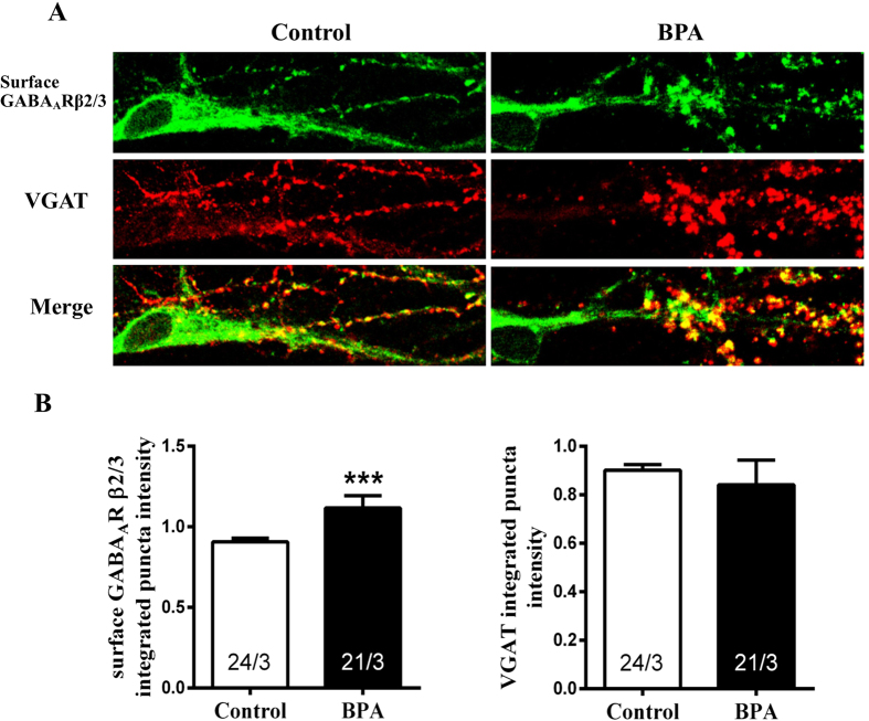
Figure 8. Effects of BPA exposure on postsynaptic surface GABAARs in cultured hippocampal CA1 neurons.
(A) Immunolabeling surface GABAARβ2/3 (green) and VGAT (red). (B) Quantification of presynaptic VGAT and postsynaptic surface GABAARβ2/3 puncta intensity. The ‘x’ of ‘x/3’ in the picture represents the number of neurons used from three independent cultures (***p < 0.001).

