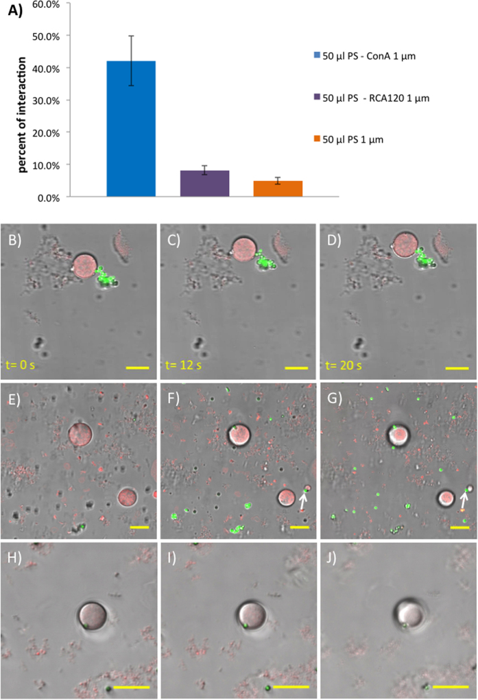
Figure 3. Glycosylated GUVs interact with lectin-functionalized PS beads through specific, multivalent sugar-lectin binding.
(A) Bar chart showing frequency of beads interacting with glyco-GUVs, from left to right: FITC-PS-Con A; FITC-PS-RCA120; FITC-PS-CO2H (RCA120 is a β-galactosyl specific lectin). (B–D) Time-lapse confocal microscopy images showing a cluster of FITC-PS-Con A beads (green) bound strongly to a glyco-GUV (red). Both the beads and the GUV move in concert. (E–J) Z-stack confocal microscopy images showing (arrows) two examples of FITC-PS-Con A beads (green) bound to the surface of glyco-GUVs (red). Inter-focal plane distances: (E,F) 3.91 μm; (F,G) 1.87 μm; (G,H) 2.10 μm; and (H,I) 3.57 μm. (B–I) are still images from videos, full versions of which are available in SI.

