Figure 7.
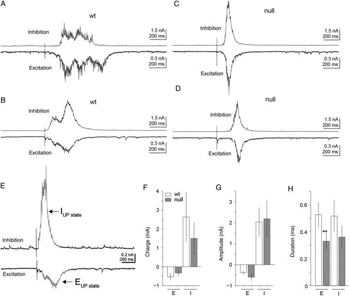
Excitatory UP-state activity was altered in Mecp2 null excitatory pyramidal layer 5 mPFC neurons. (A–D) Representative excitatory (A,B) and inhibitory (C,D) currents during electrically evoked UP-states measured within excitatory pyramidal layer 5 mPFC neurons of both Wt and Mecp2 null brain slices. (E) As in Figure 1, inhibitory and excitatory currents were electrically isolated by holding the membrane potential at either the reversal potential for excitatory (Vhold ≈ +10 mV) or inhibitory (Vhold ≈ −50 mV) synaptic currents in voltage-clamp mode. Electrical pulses delivered to the white matter elicited UP-states and either the excitatory or inhibitory components were recorded depending on the holding potential of the cell. UP-states were isolated from background based on 2-SD from baseline activity. (F–H) Summary of excitatory and inhibitory current high-conductance UP-state components of Mecp2 null (n = 14) versus Wt (n = 12) excitatory pyramidal layer 5 mPFC neurons. Across the population, the temporal dynamics of the excitatory synaptic charge was distinct in Mecp2 null neurons. (H) Overall there was a significant (P = 0.035, two-tailed Student's t-test) decrease in the mean duration of the excitatory component of the evoked UP-states in Mecp2 null neurons (mean Enull = 329 ms) compared with Wt (mean EWt = 527 ms). On average, UP-states in Mecp2 null neurons displayed more transient excitatory response dynamics compared with Wt neurons.

