Figure 1.

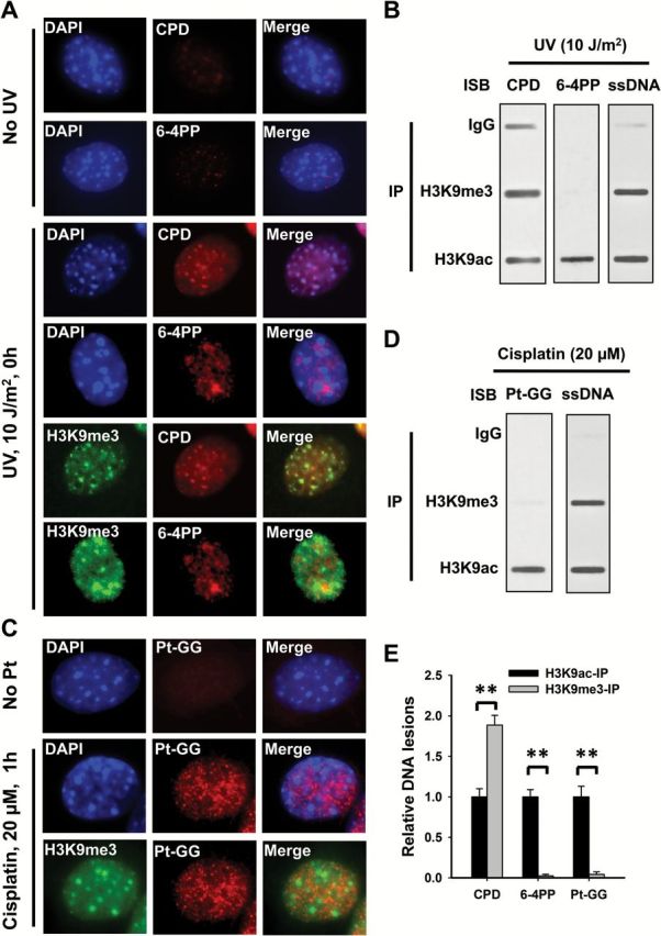
The differential formation of UV and cisplatin induced DNA lesions in chromatin. (A, C) NIH3T3 cells were UV irradiated at 10 J/m2 (A) or treated with cisplatin at 20 μM for 1h (C), or mock-treated, fixed and analyzed with mouse anti-CPD, mouse anti-6-4PP or rat anti-Pt-GG, along with rabbit anti-H3K9me3 antibodies. Detection was with cognate secondary antibodies conjugated with either AlexaFluor 594 or AlexaFluor 488. The slides were counterstained with DAPI. (B, D) Human skin fibroblast OSU-2 cells were UV irradiated at 10 J/m2 (B) or treated with 20 μM cisplatin for 1h (D), the cross-linked and sonicated DNA was subjected to the ChIP assay with either normal IgG, anti-H3K9me3 or anti-H3K9ac antibodies. The recovered DNA was subjected to the immuno-slot blot (ISB) analysis to quantitate CPD, 6-4PP or Pt-GG. ssDNA detection was used as the loading control. (E) The bands in B and D were scanned for intensity, and relative amounts of DNA lesions were calculated by first normalizing to ssDNA, then to the corresponding H3K9ac-IP samples. N = 3; Bar: SD. **P < 0.01.
