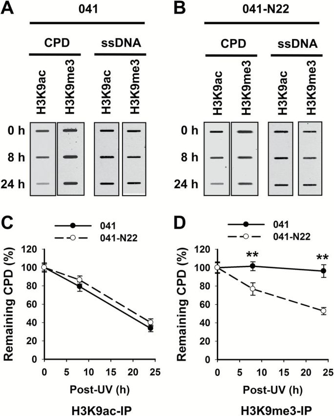
Figure 6.

DDB2 facilitates the removal of heterochromatic CPD. DDB2-deficient (041) and -proficient (041-N22) cells were UV irradiated at 10 J/m2 and further cultured for 0, 8 and 24h. The cells were fixed and subjected to the ChIP assay with either anti-H3K9ac or anti-H3K9me3 antibody. The recovered DNA was subjected to the ISB analysis to quantitate CPD. ssDNA detection was used as the loading control (A). The intensity of each band was quantitated, and relative amounts of CPD were calculated by normalizing to ssDNA first, and then to corresponding samples at 0h (B, C). N = 3; Bar: SD. **P < 0.01 compared to 041 cells at the same time point.
