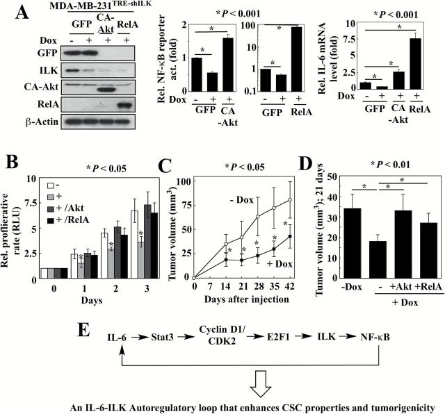
Figure 5.
Validation of the ILK-Akt-NF-κB-IL-6 signaling loop. (A) Left, confirmation of GFP, ILK, CA-Akt and RelA protein levels in MDA-MB-231TRE-shILK ectopically expressing CA-Akt, GFP-RelA or GFP, in the presence or absence of doxycycline (Dox) by Western blotting. Ectopic expression of CA-Akt and RelA reversed the suppressive effects of doxycycline-induced ILK knockdown on NF-κB luciferase reporter activity (middle) and IL-6 mRNA expression (right) in MDA-MB-231TRE-shILK cells. Data are presented as mean ± SD (n = 3). (B) Ectopic expression of CA-Akt and RelA restored the proliferation rate of ILK-knockdown MDA-MB-231TRE-shILK cells. Data are presented as mean ± SD (n = 3). +, doxycycline-treated. (C) Suppressive effects of doxycycline (Dox)-induced ILK knockdown on the growth of subcutaneous MDA-MB-231TRE-shILK xenograft tumors. Data are presented as mean ± SD (n = 6). (D) Ectopic expression of CA-Akt and RelA restored the growth of ILK-knockdown (+Dox) MDA-MB-231TRE-shILK tumors to a level comparable to that of ILK-intact controls (-Dox). Mean tumor volumes at 21 days after tumor cell injection are shown. Data are presented as mean ± SD (-Dox, n = 6; +Dox, n = 10; +Dox/+Akt, n = 10; +Dox/+RelA, n = 7). (E) Schematic diagram depicting the role of the Stat3-cyclin D1/CDK2-E2F1-ILK axis in mediating the IL-6-NF-κB feedback loop.

