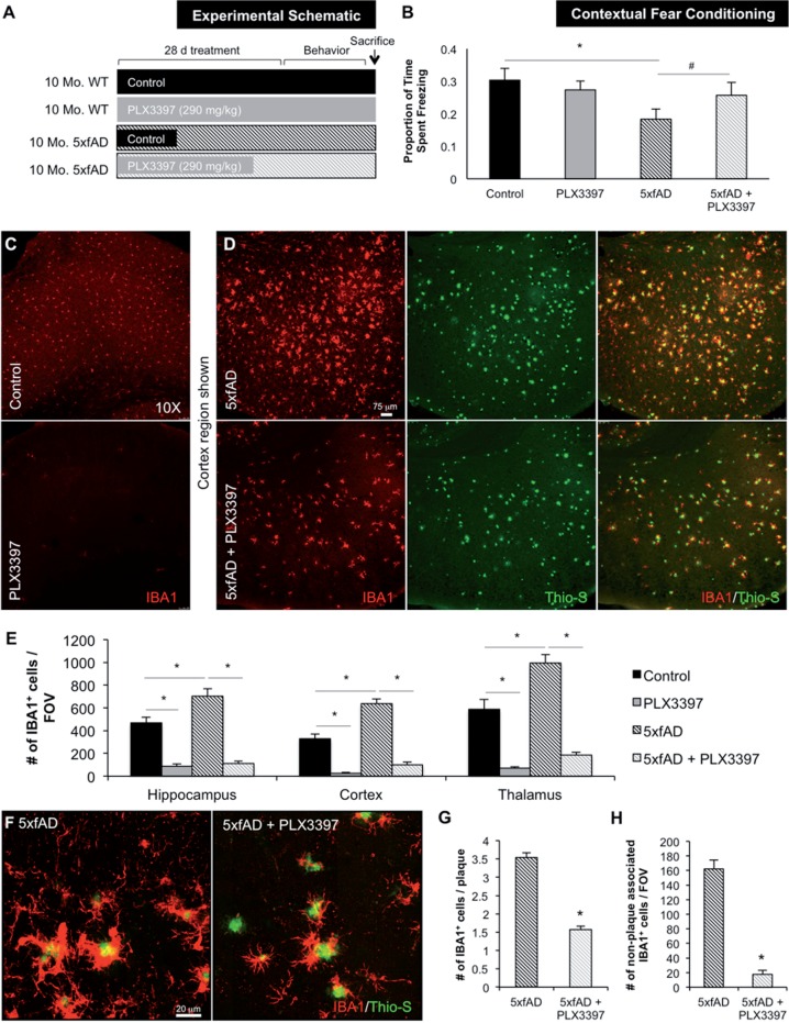
Figure 2.
Chronically activated microglia in 5xfAD mice are dependent on CSF1R signalling for their survival. Ten-month-old wild-type or 5xfAD mice were treated with control chow or PLX3397 for 28 days to eliminate microglia. (A) Experimental design. (B) In contextual fear conditioning, 5xfAD mice spent significantly less time freezing compared to control (via two-way ANOVA, P = 0.0196) and 5xfAD + PLX3397 treated mice trended to an increased freezing time (via two-way ANOVA, P = 0.0813). (C and D) Immunolabelling for microglia (IBA1 in red) and staining for dense-core plaques (Thio-S in green). (E) Microglia number is increased by ∼40% in 5xfAD mice compared to control (via two-way ANOVA, P < 0.0001). PLX3397 treatment eliminates ∼80% of microglia in both wild-type and 5xfAD mice (via two-way ANOVA, P < 0.001). (F) Representative 63X IBA1 Thio-S immunofluorescent staining of the cortex. (G) Quantification of plaque-associated microglia reveals the number of these cells was reduced by ∼50% with PLX3397 treatment (two-tailed unpaired t-test, P < 0.0001). (H) The number of non-plaque associated microglia is reduced by ∼90% with PLX3397 treatment in 5xfAD mice (two-tailed unpaired t-test, P < 0.0001). Statistical significance is denoted by *P < 0.05 and statistical trends by #P < 0.10. Error bars indicate SEM (n = 7/group).

