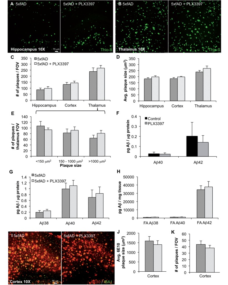
Figure 3.
Elimination of microglia does not modulate amyloid-β levels or plaque load. (A and B) Representative hippocampal and thalamic 10× images of dense core plaques (Thio-S) in 5xfAD and 5xfAD mice treated with PLX3397. (C and D) Quantification of number of Thio-S+ plaques and average area of the plaques in the hippocampus, cortex, and thalamus. (E) Microglial elimination has no effect on plaques of any size. (F) Levels of amyloid-β1–40 and amyloid-β1–42 were unchanged with microglial elimination in wild-type mice. Levels of amyloid-β1–38 were below detection threshold. (G). (H) Levels of amyloid-β species in detergent-soluble and formic acid-soluble (FA) fractions are not changed with microglial elimination. (I) Representative 10× 6E10 and IBA1 immunofluorescent images of the thalamus. (J and K) Quantification of 6E10+ plaque size and numbers reveals no effect of microglial elimination. Error bars indicate SEM (n = 7/group).

