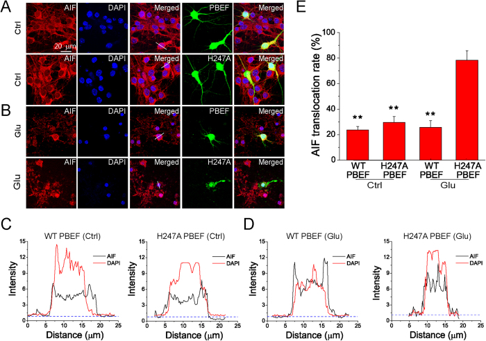
Figure 2. Overexpression of PBEF prevents AIF translocation from mitochondria to the nucleus after glutamate excitotoxicity.
(A,B) Confocal fluorescent images showing the triple staining of AIF, PBEF and DAPI in neurons under normal (A) and glutamate excitotoxicity (B) conditions. Neurons co-transfected by WT PBEF or H247A PBEF with EGFP and were treated with 100 μM glutamate together with 10 μM for 3 h. Transfected neurons were identified by EGFP. Translocation of AIF from mitochondria to nuclei is illustrated by the overlap of AIF (red) and DAPI (blue) signals. (C,D) Linescan analysis of AIF and DAPI fluorescence from the neurons indicated in (A,B). The fluorescence was normalized to the background. Notice the prevention of glutamate-induced AIF translocation by WT PBEF overexpression. (E) Summary of AIF translocation after glutamate stimulation. Data was quantified by the number of cells with overlap of AIF and DAPI among the total number of cells determined by DAPI staining. n = 3 three independent experiments. **P < 0.01 versus H247A PBEF with Glu, ANOVA test.

