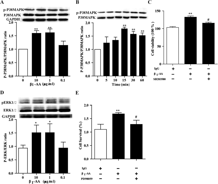
Figure 7. β1-AA promoted proliferition in cardiac fibroblast through.activating the P38MAPK and ERK1/2.
(A) CFs were stimulated with β1-AA (0, 0.1, 1, 10 μg/ml) for 30 min, then phospharylated p38MAPK and total p38MAPK were detected by western blot. (B) Immunoblot detection of phosphorylated p38MAPK (p-p38MAPK) and total VASP form CFs stimulated with β1-AA (10 μg/ml) at 0, 5, 10, 15, 30 and 60 min. (C) Cell viability of CFs were pretreated with the P38MAPK inhibitor SB203580 (1 μmol/ml) for 30 min before adding β1-AA (10 μg/ml). (D) CFs were stimulated with β1-AA (0, 0.1, 1, 10 μg/ml) for 30 min, then phospharylated ERK1/2 and total ERK1/2 were detected by western blot. (E) Cell viability of CFs were pretreated with the ERK1/2 inhibitor PD98059 (1 μmol/ml) for 30 min before adding β1-AA (10 μg/ml). Data shown here are representative of one of three individual experiments with similar results. n = 3 per group. **P < 0.01 vs. vehicle (PBS) group.

