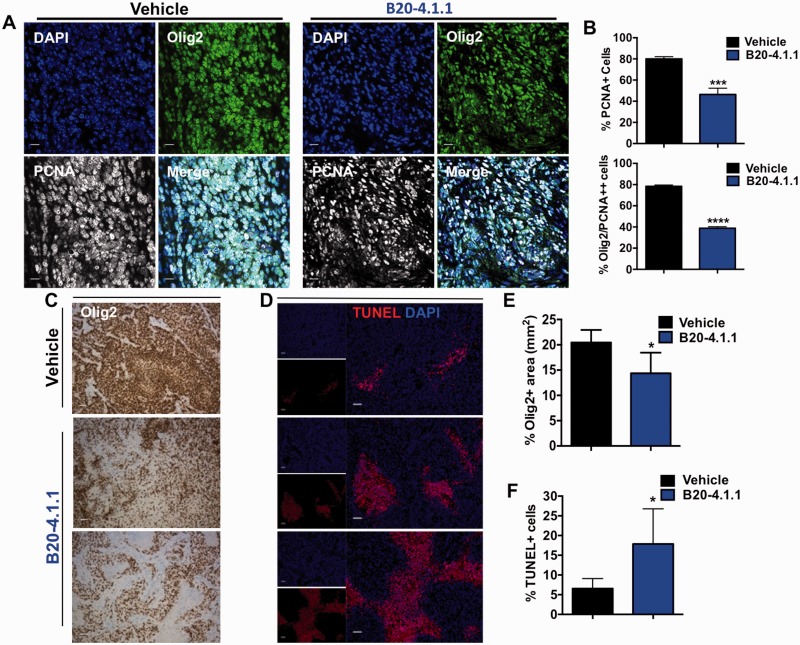
Figure 8.
B20-4.1.1 decreases proliferation and increases cell death in murine PDGF-driven gliomas. (A) Representative tumour sections and (B) quantification bar graphs of glioma proliferation using Olig2 expression as a glioma cell marker and PCNA as a proliferative marker. There was a significant reduction in the proliferation of glioma cells after B20-4.1.1 treatment. (C) Representative images of Olig2 staining in tumour sections showing decreased staining in B20-4.1.1-treated mice compared to vehicle-treated mice. (D) Representative images of TUNEL staining in vehicle- and B20-4.1.1-treated tumours and (E) corresponding quantification of Olig2-positive area (F) and TUNEL-positive cells in response to B20-4.1.1 treatment. P-values were calculated by an unpaired Student’s t-test, *P < 0.05, ***P < 0.001, ***P < 0.0001. Scale bars = 50 μm for A, C and D.

