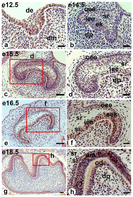
Fig. 1.
Klf10 immunostaining in embryonic mouse molar germs. a At embryonic day (E) 12.5 (e12.5), Klf10 was expressed in both the dental epithelium and the underlying mesenchyme. b At E14.5 (e14.5), Klf10 appeared in the inner and outer enamel epithelia, stratum reticulum, primary enamel knot and dental papilla. c At E15.5 (e15.5), the Klf10 expression pattern was similar to that of E14.5. e By E16.5 (e16.5), Klf10 expression was found in the secondary enamel knot, inner and outer enamel epithelia, stratum intermedium and stratum reticulum and also in the mesenchymal cells beneath the inner enamel epithelia. g At E18.5 (e18.5), positive Klf10 staining appeared in the differentiating ameloblasts and odontoblasts, the outer enamel epithelia, the stratum intermedium, the stratum reticulum and the dental papilla cells. d, f, h Higher magnification images of red-boxed areas in c, e, g, respectively (de dental epithlium, dm dental mesenchyme, oee outer enamel epithelium, iee inner enamel epithelium, ek enamel knot, dp dental pulp, sr stratum reticulum, si stratum intermedium, sek secondary enamel knot, am ameloblast, od odontoblast). Bars 20 μM (a, b, d, f, h), 50 μM (c, e, g)

