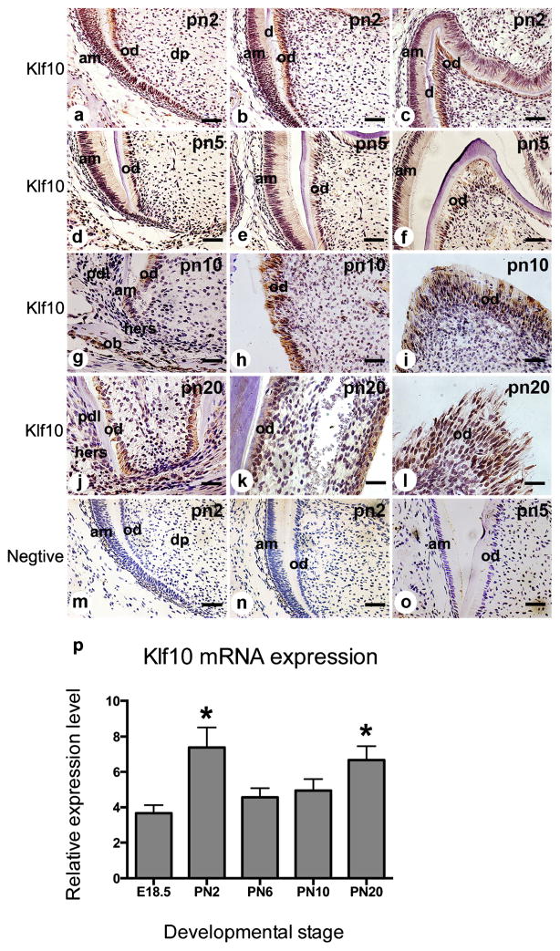
Fig. 2.
Klf10 expression in postnatal mouse molar development. a–c At PN2 (pn2), Klf10 was strongly expressed in preodontoblasts/ameloblasts and in polarizing, secretory and mature odontoblasts and ameloblasts and in several dental pulp fibroblasts. d–f At PN5 (pn5), the Klf10 expression pattern was similar to that at PN2. g–i At PN10 (pn10), Klf10 expression was apparent in ameloblasts, odontoblasts and osteoblasts, while its expression was moderately seen in dental pulp cells. Klf10 expression was barely visible in Hertwig’s epithelium root sheath (HERS) and periodontal ligament (PDL) cells. j At PN20 (pn20), Klf10 staining was observed in the odontoblasts in the root and faint staining of Klf10 was observed in HERS cells. k–l Klf10 was seen in the odontoblasts in the crown. m–o Tissue sections from PN2 and PN5 were stained with IgG as a negative control (am ameloblast, od odontoblast, ob osteoblast, pdl periodontal ligament fibroblast, hers Hertwig’s epithelial root sheath). Bars 20 μM (a–f, m–o), 50 μM (g–l). p Klf10 transcript was expressed in the first mandibular molars from E18.5 to PN20 (error bars means ± S.D.; *P<0.05 with significant difference versus E18.5)

