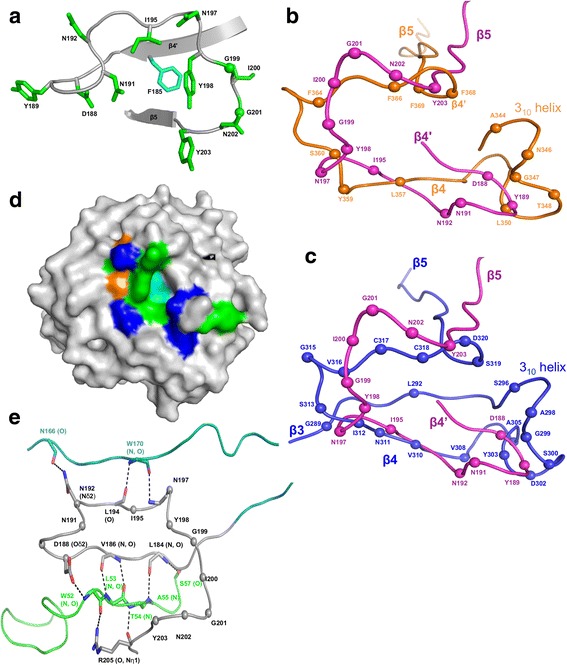
Fig. 3.

Variable Region. a Variable region of AvpA in ribbon representation. The main chain is gray and side chains of variable residues are green (green spheres correspond to glycines. The nonvariable residue F185 is shown in cyan. b Superposition of the VR of AvpA (magenta) and Mtd-P1 (orange) in Cα representation. The spheres represent variable amino acid positions. Secondary structure elements are labeled. c Superposition of the VR of AvpA (magenta) and TvpA (blue) in Cα representation. The representation is as in panel b. d Surface representation of AvpA, with variable hydrophobic residues (Y, I) green, variable hydrophilic residues (D, N) blue, variable glycines orange, and nonvariable F185 cyan. e Stabilization of the AvpA VR (gray) by insert 2 (green) and insert 4 (teal) in Cα representation. Hydrogen bonds are indicated with dashed lines
