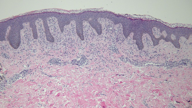
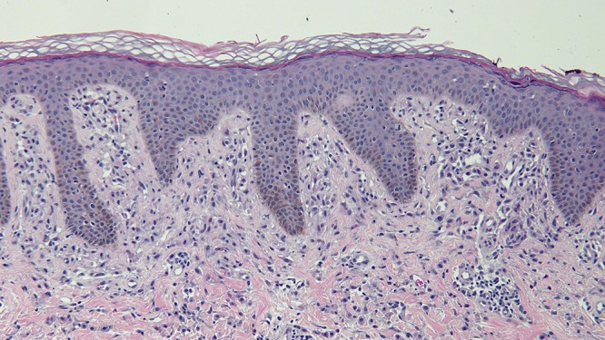
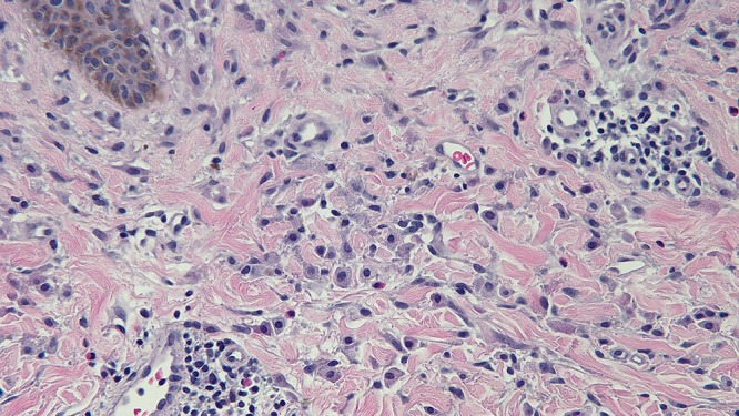
Figure 4.


Distant (a) and closer (b and c) views of the skin biopsy of the mastocytoma on the right abdomen, stained with hematoxylin and eosin, show a monomorphous mononuclear cell infiltrate in the upper dermis (hematoxylin and eosin, a = x4; b = x20; c= x 40). [Copyright: ©2016 Cohen.]
