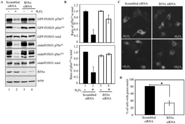
Figure 4. B55α knockdown inhibited oxidative stress-induced FOXO1 dephosphorylation and nuclear translocation.
(A) INS-1 cells were co-transfected with GFP–FOXO1 and B55α siRNA or scrambled siRNA, and immunoblotted for GFP–FOXO1 and endogenous FOXO1 using antibodies against phosphorylated (p) Thr24 and Ser256 and total FOXO1, as well as against B55α. Cells were incubated in the presence (+) or absence (−) of 100 μM H2O2 for 1 h. GFP–FOXO1 (97 kDa) is distinguished from endogenous FOXO1 (70 kDa) by its slower migration. (B) Ratios of phospho-Thr24 and phospho-Ser256 phosphorylated FOXO1 relative to total FOXO1 in untreated (−) or H2O2-treated (+) cells transfected with scrambled or B55α siRNA. Ratios were normalized to untreated cells transfected with scrambled siRNA. Band intensities were acquired from (A) using Bio-Rad quantity analysis software and represent the means±S.D. of four samples. (C) INS-1 cells were co-transfected with GFP–FOXO1 and B55α siRNA or scrambled siRNA. GFP localization was visualized by fluorescence microscopy in untreated and H2O2-treated cells. (D) Percentages of GFP-positive nuclei after H2O2 treatment in scrambled siRNA or B55α siRNA-transfected cells. At least 150 cells were counted in three independent experiments. *P <0.01.

