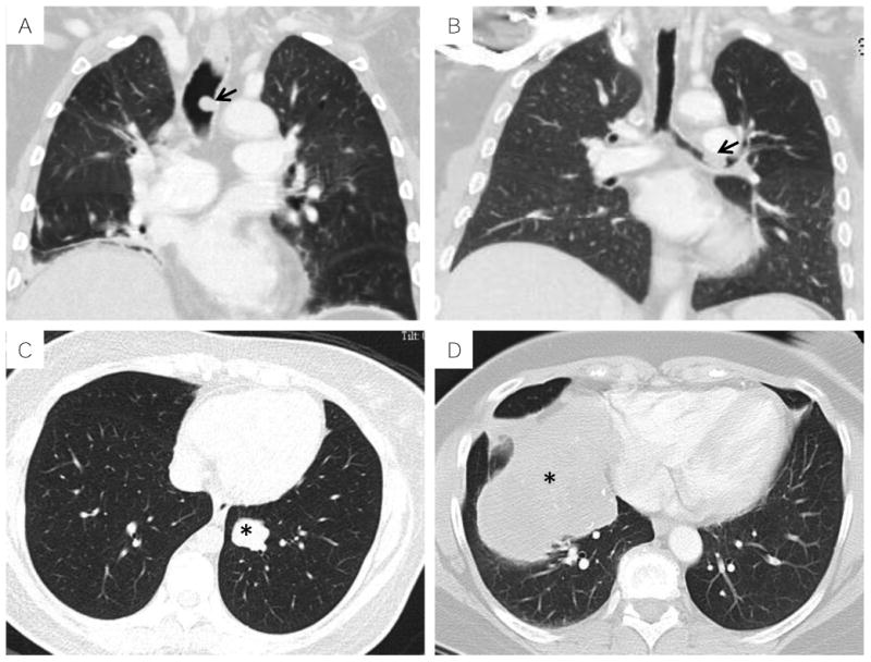
Figure 1.

Radiologic features of thoracic MTs on computed tomography: A, sessile endotracheal polyp (arrow, Case #5); B, sessile endobronchial polyp (arrow, Case #8); C, well circumscribed intraparenchymal nodule (asterisk, Case #6); D, large intraparenchymal mass with calcifications and mass effect (asterisk, Case#3).
