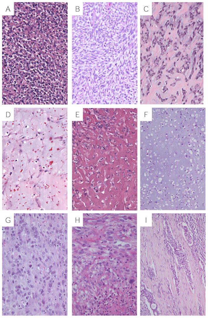
Figure 4.

Histologic features of thoracic MTs: A, sheets of round cells with clear cytoplasm (Case #3); B, sheets of spindle cells with clear cytoplasm (Case #3); C, plasmacytoid cells in a reticular pattern (Case #8); D, spindle cells in a myxoid stroma (Case #2); E, epithelioid cells in a hyalinized matrix (Case #6); F, clear cells in a chondroid matrix (Case#3); G, epithelioid cells with moderate cytologic atypia (Case #7); H, Mitotically active spindle cells with focal necrosis (Case#1); I, Myoepithelial tumor cells within lymphovascular channels (Case #2).
