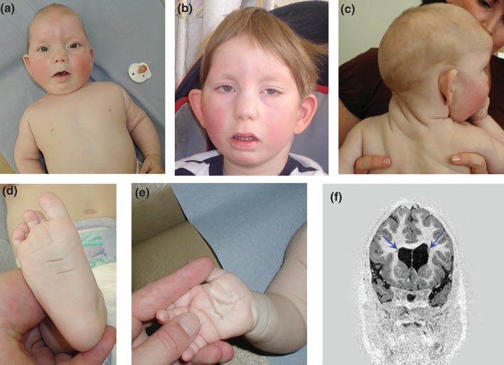
Figure 1.

(a, b) Proband at ages 7 months and 8 years, respectively. Note the prominent metopic ridge, turricephaly, sparseness of lateral eyebrows with medial flare, long palpebral fissures, eversion of lower eyelids, low‐set prominent ears. (c) Thick, wrinkled skin on the neck posteriorly. (d, e) Deep plantar and palmar creases with fetal finger pads. (f) Coronal MRI image from inversion recovery volume sequence demonstrating two grey matter rounded nodules (blue arrows) adjacent to the borders of the lateral ventricles, in keeping with periventricular heterotopia. Written consent from the family to publish these images was obtained.
