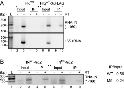
Figure 7.

Hfq‐RNA‐IN immunoprecipitation (RIP) assay.
A. hfq− cells containing a chromosomal IS 10 HH104‐kan element (DBH337) were transformed with plasmids expressing HfqWT (pDH904) or HfqWT‐3xFLAG (pDH909; C‐terminal 3xFLAG tagged Hfq). Hfq was immunoprecipitated (IP) from cell lysates with ANTI‐FLAG ® M2 magnetic beads; untagged Hfq (HfqWT) served as a negative control. The first 160‐nt of RNA‐IN (top panel) or nts 1071–1425 of 16S rRNA were detected by RT‐PCR (see Experimental procedures). Samples were analyzed on a 2% agarose gel that was stained with ethidium bromide. No reverse transcription (−RT) controls are shown (lanes 2, 4, 6 and 9). L is a DNA ladder (lane 1).
B. hfq− cells (DBH337) were co‐transformed with HfqWT‐3xFLAG plasmid and a plasmid encoding either WT or M5 transposase‐lac Z. Hfq RIPs were performed as in A. Band intensities for the input and IP RT‐PCR signal were quantified with an AlphaImager 3400 (Alpha Innotech).
