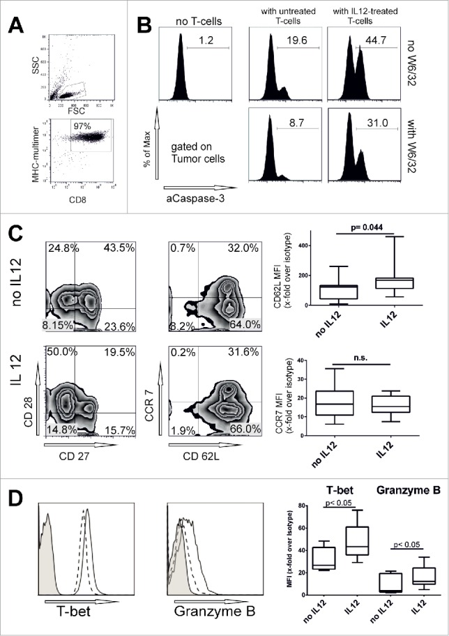
Figure 2.

IL12 enhances antigen-specific cytotoxicity and retains CD62L expression. (A) Representative dot plots with gating strategy for Melan-A-MHC-multimer purified T cells. (B) MHC-multimer-purified Melan-A sp T-cells were cultivated with or without IL12 (10 ng/mL) for 48 h and then used in a caspase-3 apoptosis assay against the HLA-A02:01+ melanoma cell line FM55 (without exogenous peptides). HLA-ABC blocking antibody W6/32 (10 µg/mL) was added to the tumor cells 15 min before the T cells were added and was present during the assay. After 4 h tumor cells were stained for activated caspase-3 (for the gating strategy see Fig. 3B). E/T-ratio 2:1. Mean and SD from three independent experiments. (C) Melan-A sp. T cells in the proliferative phase were incubated for additional 48 h with or without IL12 and analyzed for various surface markers (gated on CD8+ MHC-multimer+ T cells as shown in Fig. 2A). Right panels: Summary of ten experiments, depicted as change in MFI (x-fold over isotype, mean and quartiles). For additional results see Fig. S1E. (D) Representative histograms of T-bet- and Granzyme B-expression. Filled: isotype, dashed line indicates no IL12, continuous line with IL12 treatment. Right: Summary of more than five experiments as MFI results (x-fold over isotype) are from more five experiments, including clonal cell populations (STEAP1, PRAME and clonal Melan-A cells). For all experiments in C and D: box, line and whiskers indicate: 25th to 75th percentile, median and min. to max. Significance was corrected for multiple comparisons.
