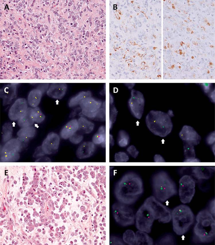
Figure 2.
Heterozygous EWSR1 deletion in SMARCB1-deleted tumors. A: Myoepithelial carcinoma composed of relatively monotonous epithelioid to round cells arranged in cords in a collagenous stroma (Case 3, 400x) showing (B) focal reactivity for cytokeratin (left, 400x) and S100 (right, 400x). The 3-color FISH revealed homozygous SMARCB1 deletions (lack of both red signals) with heterogeneous EWSR1 abnormalities, including (C) heterozygous deletions (arrows: one allele retained the paired yellow-and-green signals but with diminished yellow intensity, indicating partial deletion of centromeric part, while the other allele showed loss of green signal, consistent with deletion of telomeric part) and (D) break-apart (arrows: yellow-and-green split signals of smaller size, in keeping with concurrent deletions of centromeric and telomeric parts outside the EWSR1 gene locus). E: Proximal-type epithelioid sarcoma with myxoid changes and rhabdoid morphology (Case 4, 400x). F: The 3-color FISH showed heterozygous SMARCB1 (one retained red signal) and EWSR1 regional deletions (arrows: the involved allele containing a tiny residual green signal in keeping with a larger deletion across centromeric and telomeric part). [Color figure can be viewed in the online issue, which is available at wileyonlinelibrary.com.]

