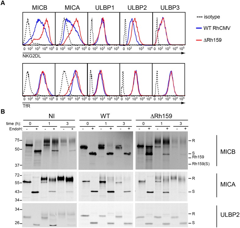
Fig 5. Deletion of Rh159 rescues intracellular transport and surface expression of MICA and MICB upon RhCMV infection.
A) Comparison of NKG2DL surface expression upon infection with RhCMV or ΔRh159. U373-NKG2DL cells were infected with RhCMV (blue) or ΔRh159 (red) (MOI = 3) for 48 h. Cell surface levels of NKG2DL or TfR were determined by flow cytometry, using specific antibodies and compared to isotype control (dotted). Depicted is NKG2DL or TfR surface expression on infected cells gated for RhCMV IE2+ expression. The results shown are representative of three or more independent experiments. B) Biosynthesis and maturation of NKG2DL in uninfected U373-NKG2DL cells or upon infection with RhCMV or ΔRh159. U373-NKG2DL cells were uninfected (NI), infected with RhCMV (WT) or ΔRh159 (MOI = 3) for 24 h, verified by light microscopy as having 100% CPE, then metabolically labeled with [35S]cysteine and [35S]methionine for 30 min prior to chasing the label for the indicated times. The indicated NKG2DLs were immunoprecipitated from cell lysates with specific mAbs. Immunoprecipitates were split and digested with EndoH (+) or mock treated (-) then analyzed by SDS-PAGE and autoradiography.

