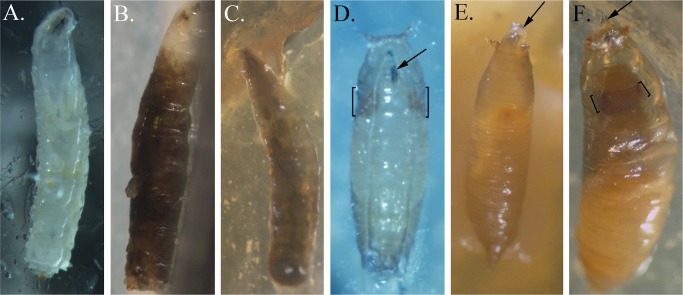
Fig 6. Larval and pupal phenotypes of D. melanogaster fed with strains of Pseudomonas spp.
(A) Ventral view of a third instar control larva showing normal clear hemolymph and internal organs. (B) Side view of a dead SBW25-fed third instar larva showing systemic melanization of the hemolymph. (C) Dorsal view of a dead BG33R-fed third instar larva with complete melanization of the hemolymph. (D) Ventral view of a mid-stage control pupa showing normal extrusion of the mouthparts (arrow) and normal size of pupal eyes (brackets). (E) Dorsal view of a dead SS101-fed pre-pupa with extended mouthparts (arrow) and no head involution. (F) Dorsal view of a dead A506-fed pharate adult with extruded mouthparts caught within the pupal case (arrow). The head and eyes (brackets) are smaller and more recessed than in normal pupae.

