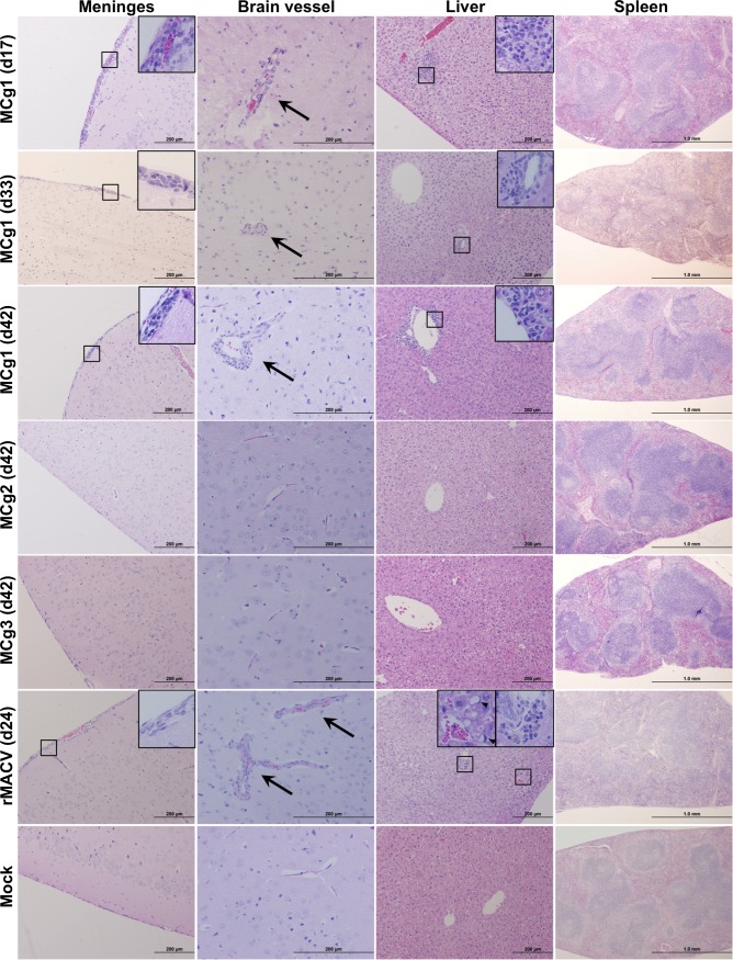
Fig 4. Histopathological changes in the brain, spleen, and liver from infected mice at 17 dpi, the terminal stage and 42 dpi.
Meningitis and perivascular cuffing (Arrow) were observed in the brains of MCg1-infected mice at 17 dpi, the terminal stage (33 dpi), the end of the study (42 dpi) and the brain of rMACV-infected mice at the terminal stage (24 dpi). Periportal infiltrates or focal inflammation were present in the livers of MCg1-infected animals at 17 dpi, 33 dpi and 42 dpi. Periportal infiltrates and microvesicular steatosis (Arrowhead) were observed in the liver of rMACV-infected animal at 24 dpi. Reactive white pulp hyperplasia was mildly disturbed in the spleen of MCg1-infected animal at 33 dpi and moderately in the spleen of rMACV-infected animal at 24 dpi. No significant histological change was observed in MCg2- and MCg3-infeceted animals at 42 dpi. Magnifications, x4 (Spleen), x10 (Meninges and Liver), x20 (Brain Vessel) and x40 (insets).

