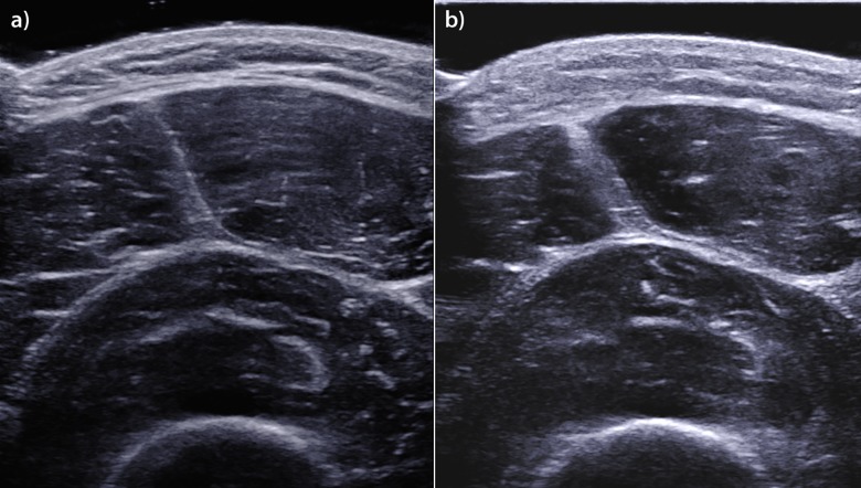
Fig 4. Edematous and architectural thigh muscle changes at the Finish assessment.
US images of the quadriceps femoris muscle, which were obtained in the axial plane during the Pre session (a) and the Finish session (b). Note the edematous thickening of the skin and the muscle fascia in the image obtained during the Finish session.

