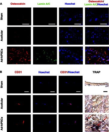
Fig. 5. hiPSC-derived osteoblasts contributed to the repair of critically sized cranial defects through the formation of vascularized neobone tissue.

(A and B) Immunofluorescence staining for osteocalcin (red) and human-specific lamin A/C (green) (A) and CD31 (red) along with nuclei (blue; Hoechst), as well as TRAP staining (B) of cranial bone defects treated with Ad-hiPSC–laden (Ad-hiPSCs) and acellular matrices, and sham group after 16 weeks of implantation. Red arrows indicate TRAP-positive stains. Scale bars, 50 μm.
