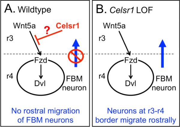
Figure 7. Model of Celsr1 function in repressing rostral migration of FBM neurons.
We propose that Wnt5a, which is expressed along the midline in r3 and more anterior regions of the hindbrain, can act as a chemoattractive cue to facilitate the migration of FBM neurons located in anterior r4, adjacent to the r3/r4 boundary, rostrally into r3 and r2. A, In a wild type hindbrain, Celsr1 expression in the ventricular zone inhibits Wnt5a-dependent activation through an unknown mechanism (?), blocking the rostral migration of FBM neurons into r3. B, In a Celsr1-deficient hindbrain, the inhibition is relieved and FBM neurons migrate rostrally into r3 toward the Wnt5a source.

