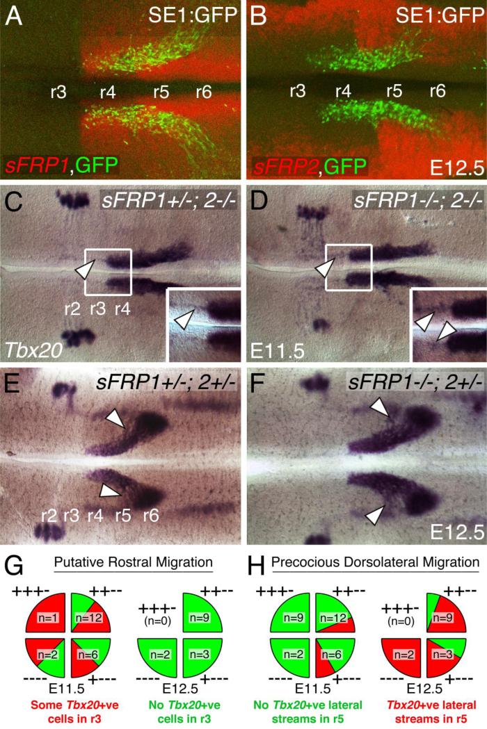
Figure 8. Wnt antagonist genes sFRP1/2 modulate caudal migration of FBM neurons.
A-F, Flat-mounted hindbrains (left is rostral) processed for ISH with sFRP1 (A), sFRP2 (B) and Tbx20 (C-F). The distribution of GFP-expressing FBM neurons in A, B (from SE1::GFP embryos) helps to delineate rhombomere boundaries. A, At E12.5, sFRP1 is strongly expressed caudal to the r3/r4 boundary in the entire neuroepithelium. Although not apparent in fluorescent ISH, sFRP1 is also expressed weakly in r3 and r2. B, sFRP2 is weakly expressed in medial regions, and strongly expressed along the lateral margins of the hindbrain at all axial levels (r3-r6). C-D, In E11.5 hindbrains, when 3 or more copies of sFRP1/2 are removed, a small number of Tbx20-expressing cells (arrowheads) appear to migrate rostrally into r3 (see insets). There are no defects in the caudally migrating streams of FBM neurons. E-F, In E12.5 hindbrains, when 2 or more copies of sFRP1/2 are removed, the caudal migration streams frequently contain ectopic streams indicating precocious dorsolateral migration (arrowheads). Interestingly, there are no rostral migration defects. G-H, Summary data showing distribution of migration defects among various sFRP1/2 compound mutants at E11.5 and E12.5. The four quarters in each “pie” show the breakdown of two phenotypes (red and green sectors) in the indicated sFRP1; sFRP2 genotypes. For example, ++−− corresponds to sFRP1+/+; sFRP2−/−. G, While over 50% of E11.5 mutants (12/21 embryos) contain some rostrally migrating Tbx20+ve cells, these cells are no longer evident in E12.5 mutants (14/14 embryos). H, While less than 20% of E11.5 mutants (5/29 embryos) contain precocious lateral streams of Tbx20+ve cells, most E12.5 mutants (11/14 embryos) contain such streams.

