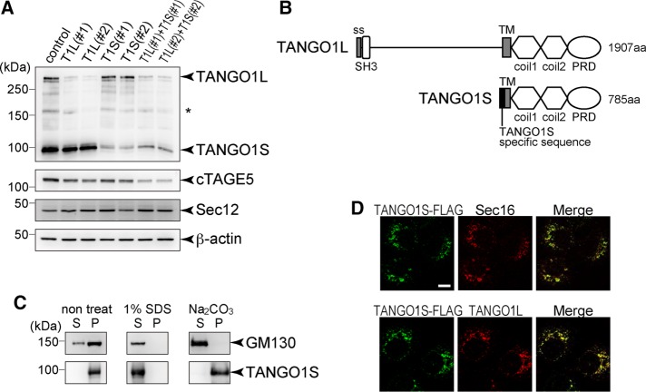
FIGURE 1:
TANGO1S is an integral membrane protein localized at ER exit sites. (A) HSC-1 cells were transfected with the indicated siRNA(s). After 72 h, proteins were extracted and subjected to SDS–PAGE, followed by Western blotting with anti–TANGO-CC1, cTAGE5, Sec12, and β-actin antibodies. *Nonspecific cross-reaction. (B) Schematic representation of human TANGO1L and TANGO1S domain organization. coil, coiled-coil; PRD, proline-rich domain; ss, signal sequence; TM, transmembrane. (C) Membrane fractions of HSC-1 cells were untreated or extracted with SDS or sodium carbonate, followed by centrifugation to separate the solubilized fractions (S) from the pelleted fractions (P). Both fractions were subjected to SDS–PAGE and Western blotting with anti-GM130 or anti–TANGO1-CC1 antibody. (D) TANGO1S-FLAG expression was induced by incubation with 10 ng/ml doxycycline for 24 h in an HSC-1 stable cell line. Cells were fixed and costained with anti-FLAG antibody and anti-Sec16 or anti-TANGO1L antibody. Scale bar, 10 μm.

