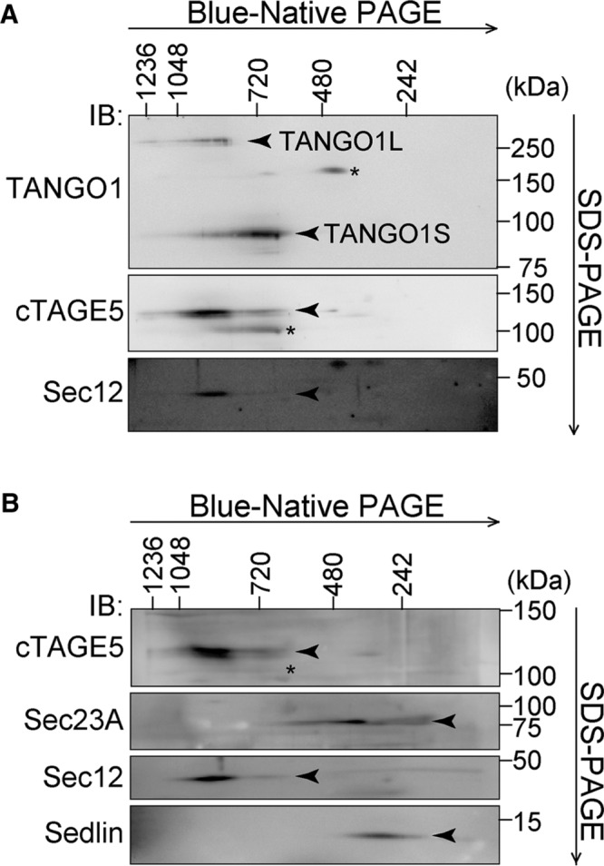
FIGURE 4:

Two-dimensional Blue-Native PAGE/SDS–PAGE analysis of HSC-1 cell lysates. HSC-1 cell proteins extracted with 1% digitonin were subjected to 4–15% Blue-Native PAGE in the first dimension and SDS–PAGE in the second dimension, followed by Western blotting with (A) anti–TANGO1-CC1, anti-cTAGE5, and anti-Sec12 antibodies or (B) anti-cTAGE5, anti-Sec23A, anti-Sec12, and anti-Sedlin antibodies. *Nonspecific cross-reaction.
