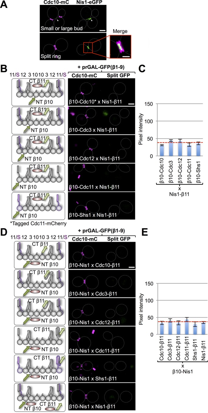
FIGURE 6:

Bud neck–localized protein Nis1 does not interact directly with septins. (A) Cells (strain GFY-42) expressing Cdc10-mCh and coexpressing Nis1-eGFP under the control of its endogenous promoter from a CEN plasmid (pGF-IVL553) were grown and visualized as in Figure 4A. Top, GFP signal (white triangles) adjacent to the septin collar in budded cells before cytokinesis. Bottom, during cytokinesis, Nis1-eGFP localizes between the two rings generated by splitting of the septin collar. Inset, enlarged view of the merged image; scale bar, 1 μm. (B) Diploids (128, 121, 142, 135, and 149) expressing Nis1-(linker)33-β11 and the β10-(linker)18–tagged versions of each of the five mitotic septins (left), as well as either Cdc10-mCh or Cdc11-mCh (as indicated), visualized as in Figure 1B (right). (C) Quantification, as in Figure 1C, of the data in B. (D) Diploids (157–161 and 163) expressing β10-(linker)18-Nis1 and C-terminally (linker)33-β11–tagged versions of each of the five mitotic septins (left), as well as Cdc10-mCh, were visualized as in Figure 1B (right). (E) Quantification, as in Figure 1C, of the data in D.
